This week was spring break so I went to Florida. I actually did work on my test corrections though so that was kind of productive. Senior year is coming to and end and the motivation is increasingly dimming so I guess next week I will try to work and staying focused and finishing strong.
3 ques 2/28
This week we took our test on the endocrine system and I actually did fairly well. Keeping up with my notebook really helped and taking all the notes. Next I will try to continue to do well on the next unit by finishing all the coloring plates and taking notes.
3 questions 1/30
This week we talked about neurons and went over our leech lab where we looked at a simple nervous system. We also got new coloring plates on different types of the nervous system and completed a blog on sports drinks and the longest nerve in the body. Next week we are reviewing and taking our test and I’m am going to study for it and complete my glogs!
The Structure of a Neuron Essential Questions
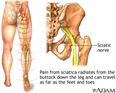
Why do athletes use sports drinks? Sports drinks replenish your electrolytes after and during exercise. Those sports drinks contain electrolytes such as chloride, sodium, and potassium. These electrolytes are lost from the body through sweat and potassium and sodium play a huge role in muscle contractions. The sodium potassium pump is a significant step in a muscle correction because it controls the ions traveling on each side of the cell membrane. When those ions are lost through sweat the sports drinks can replenish them thus continuing the ability to produce strenuous exercise with muscle contractions. The longest nerve in the body is the sciatic nerve, and it runs from the bottom of the spinal cord to the big toe on each foot. That is a length of approximately three feet for one neuron!

Leech Lab
The point of the leech lab was to view a simple nervous system in a leech because even simple nervous systems perform much of the same functions as the complex ones, as we discuss in anatomy. I reviewed that the basic unit was the neuron and that was enforced in the explanation and use of electrode stimuli. I also got to identify five different cell types that were stimulated by the different utensils. These cells were named N,T,P,R, and X. This lab is applicable to people because although leeches are simple creatures the anatomy of their nervous system is very similar to that of humans. These simulations can help represent the human nervous system because most nerve cells are stimulated similarly. In the picture shown is an example of a dyed cell from the leech lab. Seeing it in UV light was really interesting and depicted what the cells looked like.
My source was the hhmi leach lab

Play doh brain!
The human brain is a very strangely structured piece of art. You have the diencephalon which sits on the brain stem and is enclosed by cerebral hemispheres and is made of the Thalamus, Hypothalamus, and Epithalamus. The epithalamus forms the root of the third ventricle. It also houses the pineal body. The thalamus is what I like to refer as the “sensory switchboard.” It surrounds the third ventricle and acts as a relay station for sensory impulses. It then transfers those impulses to the correct part of the cortex for localization and interpretation. Lastly in this part of the brain we have the hypothalamus. This part of your brain plays a HUGE role in the “fight or flight” experience. It is located under the thalamus and is important in controlling body temperature, water balance, and metabolism as well as playing a big role in the limbic system of emotions. Now, in the brain stem you have the midbrain, which acts as a reflex center for vision and hearing and has two bulging fiber tracts called the cerebral penduncles. Next, is the medulla oblongata which contains important control centers such as heart rate, blood pressure, breathing, swallowing, and vomiting. The medulla is the lowest part of the brain stem. Then of course there is the pons which is a bulging center part of the brain stem, is mostly composed of fiber tracts, and includes nuclei involved in the control of breathing. The reticular formation is next and it is involved in motor controls of visceral organs. the RAS plays a role in the awake and sleep cycle. You can thank it for your circadian rhythm later. Last in the brain stem is the cerebellum which is also know as the “mini brain.” It is composed of two hemispheres and provides involuntary coordination of body movements. Then you have the specialized areas of the brain like the somatic sensory area with receives impulses from sensory receptors, or the motor area that controls the impulses being sent to skeletal muscles, or even the Broca’s area which is involved in our ability to speak. All of the different parts of the brain must perform a different task to keep our body working the way we like it to. If something were to happen to the medulla for example, living would be nearly impossible. Each part of the brain plays a vital role in taking care of our body. We need each part of the brain to function so the body can keep doing all the things we like to do! So thank your Broca’s area the next time you’re feeling chatty, or even your hippocampus the next time you remember some funny memory!
For my sources I would like to cite my anatomy notes on the brain as well as my psychology notes on the brain!
Here is a picture of my play doh brain.


3 questions 1/15/15
This week we graded our test on the muscular system. I learned I need to study more of the microscopic reactions and fully read each question. Then we did test correction on each question! To more prepare for class I should probably take the notes and pay more attention to them! Next I will try to read all the questions thoroughly especially since we are in the nervous system and it’s very complex.
Skeletal Muscle!
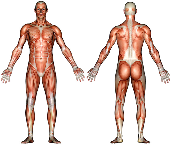
Okay, here we go with a microscopic look at the skeletal muscle. First the skeletal muscles in a human body are about 40% of the body weight according to my coloring packet. Inside the skeletal muscle you have a bundle of fibers. they are cylindrical striated cells called fibers. Next, is the cell, or muscle fiber. These can be very long because they extend from one bone to another. inside the muscle fibers is the myofibrils which are hundreds of banded cylindrical myofibrils that run the length of each cell. So this skeletal muscle is pretty much like a giant puzzle. Then of course you have actin filaments that are the proteins needed in the cell. You also have myosin filaments which are assemblies of protein molecules to make myosin. The contractile elements in the skeletal muscle are the A band, Thick filaments, I band, thin filaments (actin filaments), H zone, z line, and sacromere. The bands are responsible for the striations in the cell look. The thin filaments run vertical through the cell and the thick ones peripheral. They interact through the cross bridges which look like buds or little clams. When a muscle contracts the I band shortens and that makes the H zone shorten. Notice the thick and thin filaments do not change which means they never change length. So how exactly does it contract and where does it get the energy? For skeletal muscle fibers to contract: The fiber must be stimulated by a nerve ending, an action potential must be generated along the sarcolemma, the action potential must be propagated along the sarcolemma, and intracellular calcium levels must rise to trigger contraction. When the ACh is release it travels down the t tubules then Ach is broken down by acetylcholine esterase, which terminates stimulation of the sarcolemma. If we did not have muscle movements it could mean that we would never make our bodies warm or we would never be able to move! Some of the roles also include food digestion and the aid in blood flow so without the muscle system we would not be alive!

If you are interested in learning more visit:
http://srufaculty.sru.edu/timothy.smith/tds-web-pages/TDS-web-atlas/Microscopic%20Anatomy%20of%20Skeletal%20muscle.htm
My sources:
Class notes
Class coloring packet
google images
3 question 11/21
This week we took a test on bones. I did terrible and cried. Then we did test corrections and I still didn’t know it so I cried some more. This was all in class let me remind you. Next week I will not be in class Because I have an interview for a medical program! Hopefully I don’t cry. Then it’s thanksgiving so I will be eating lots of turkey and gaining adipose tissue.
Skeletal System Blog
Okay so we just covered the skeletal system. In essence, this systems function is the framework for the entire body. It protects the internal organs. For example the rib cage protects many vital organs like the heart and lungs. Without the rib cage protecting those organs they may be more susceptible to damage caused by rigorous motions. The integumentary system also works in protecting the body. Basically this system is an organ all in itself– the skin! The integumetary system protects the body by keeping toxins out, blood in, working as pain receptors, keeping temperature constant, and much more! This system protects the body physically, (as a barrier for scrapes etc) but also chemically (water balances, sweat, and mineral balance). Along with both of these systems protecting the body also comes the instability that happens with imbalance. If the systems happen to be imbalance many diseases can happen. For example, osteoporosis for the skeletal system, and herpes for the integumentary system!
http://i.livescience.com/images/i/000/036/339/i02/human-body-skeletal-130120-1.jpg?1359999736
https://sp.yimg.com/ib/th?id=HN.608015971869590006&pid=15.1
In case you need help with each system here are some links!
http://www.faqs.org/health/Body-by-Design-V1/The-Integumentary-System.html
http://www.livescience.com/22537-skeletal-system.html
My sources:
Class notes!
virtualgardner2.weebly.com


